Prof. Dr. Tansel Ansal Balcı, Head of Nuclear Medicine Department of Fırat University Hospital, gave information about PET-CT examination.
Prof. Dr. Tansel Ansal Balcı said that PET-CT is a diagnostic method in which fusion images are created by combining nuclear medicine and anatomical imaging and can be used in most cancer diseases, some neurological diseases and even some infectious diseases. Prof. Dr. Balcı emphasized that PET-CT is very important in cases where cancer is suspected, after cancer diagnosis, whether the cancer has spread to any part of the body, whether the patient responds to the treatment in patients who have undergone treatment, and whether the cancer has recurred. Stating that they can visualize the areas where the cancer is located with the radioactive substances they give to their patients, Prof. Dr. Balcı noted that the cancer tissue shows where it is located by luminescence through radioactive labeled substances given intravenously, and that thanks to the PET-CT device, they can easily see where the cancer is located and thus they can obtain very impressive images. Prof. Dr. Balcı said that they usually use a sugar-like molecule marked with a radioactive substance and that they give this substance to the patient intravenously before the examination and then this substance is placed in the cancerous tissue in the body, so that cancerous areas can be detected more clearly. Prof. Dr. Balcı stated that PET-CT is not a screening test, but this test can be performed in people with very strong suspicion of cancer, and that PET-CT test has become frequently used especially in dementia patients, and that it is very effective in terms of diagnosing and determining the type of dementia. Prof. Dr. Balcı stated that before the PET-CT procedure, especially patients with a diagnosis of diabetes should pay attention to their diabetes, since they give the patient a radioactive substance containing a similar sugar molecule, the blood sugar level of the patients should not be high during the examination in order to obtain the best image from the patient and to interpret the resulting image in the best way.
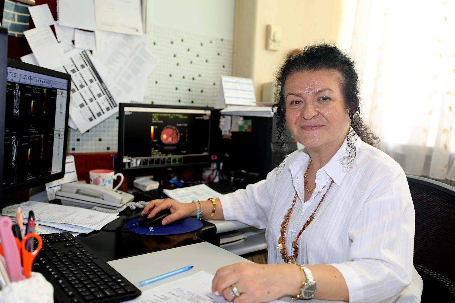
Prof. Dr. Tansel Ansal Balcı stated that PET-CT images are examined very carefully and meticulously, compared with old images, if any, and the film is reported after examining issues such as whether the patient has cancer, whether the existing cancer has spread, where it has spread if it has spread, whether the cancer has recurred if there is a previous history of cancer, whether the cancer has recurred again, and whether the treatment is beneficial or not. He explained that the treatment of the patient is organized after the written report reaches the doctor requesting the examination.
Stating that PET-CT scans have been performed at Fırat University Hospital for about 1 year, Prof. Dr. Balcı said that they had been waiting for this device for many years, and that the PET-CT device was brought to the hospital and the university thanks to the visionary work of Fırat University Rector Prof. Dr. Fahrettin Göktaş.
İHA Haber Kodu: 20241107AW324449